This post was written based on a presentation by Jonathan Shirazi, MD, FACC, FHRS, PPG – Cardiology.
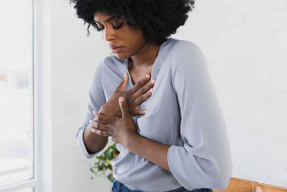
A heart palpitation is a fluttering sensation felt in the chest or neck that can be from a rapid, strong or irregular heartbeat. Sometimes, these abnormalities are triggered by our heart, sometimes our nerves, and sometimes neither. What follows are some of the many causes of palpitations and some context to help understand the variation in treatment recommendations. Some of the data presented is a bit dated, but still serves as a good starting point to understanding more about palpitations.
Instances
Palpitations are common. Approximately 6 to 11% of people report experiencing palpitations over the course of a year. In a survey of patients visiting their primary care providers, 16% of patients have complained of a fluttering sensation. In fact, palpitations are the second most common reason patients are referred to a cardiologist.
Causes
Of 190 people with palpitations followed for one year, the sensation was attributed as follows:
- 43% were caused by cardiac causes
- 31% were caused by anxiety or panic disorder
- 6% were caused by medications or supplements
- 4% were caused by other non-cardiac causes
- 16% of the time, no cause was found
Regarding “non-cardiac causes” fluttering in the chest can be caused by muscle spasms of the intercostal muscles, serratus anterior muscles, or even pectoralis minor. Only rarely is the GI tract the culprit; in those rare cases, esophageal spasm is the diagnosis causing symptoms.
When we look at medications and supplements that can trigger palpitations, the most common sources are:
- Alcohol (acute use, chronic use, or withdrawal from chronic use)
- Nicotine (in any form)
- Too much Caffeine
- Decongestants
- Bitter orange, elderberry, ephedra, hawthorne, guarana, valerian and many more supplements
- Marijuana, amphetamines and cocaine
Avoiding or limiting these substances can be helpful for most patients feeling fluttering sensations.
The second most common cause of palpitations is anxiety and panic attacks. An estimated 19.1% of American adults report an anxiety disorder annually, and approximately 31.1% of U.S. adults experience an anxiety disorder at some time in their lives. With the prevalence of anxiety and panic disorders so high, it is no surprise that a large number of fluttering events are attributed to the condition.
The role of the heart rhythm
When we start investigating potential cardiac issues, heart rhythm is key. We have to identify the heart rhythm at the time the patient experiences palpitations. We might capture this using:
- ECG (ECG at time of symptoms)
- Electrophysiology study
- Event monitor (1-4 weeks)
- Holter (24-48 hours)
- Smartwatches with ECG features (e.g. AppleWatch gen 4 or earlier, Samsung Watch, etc.)
- AliveCor Kardia Mobile or other single lead home ECG capture devices
If the patient doesn’t experience symptoms when we’re monitoring them, it may be worthwhile to continue to monitor longer (see loop recorder commentary, below). The key is that the patient has to feel the palpitations when the heart rhythm is being recorded in order to rule in or rule out an arrhythmia as the cause.
Screening with echocardiogram
Patients with recurrent palpitations, or patients with palpitations and other potential cardiac symptoms, typically warrant an echocardiogram.
An echocardiogram is an ultrasound of the heart that provides information regarding:
- Heart size
- Heart function
- Valve function
While rare, it’s still important for a cardiologist to evaluate the patient for cardiac abnormalities such as hypertrophic cardiomyopathy and mitral valve prolapse.
Here are several arrhythmic causes of palpitations. This is certainly not all encompassing and these are brief highlights of each arrhythmia.
Premature ventricular complexes
Premature ventricular complexes (PVCs) are extra heartbeats that disrupt the regular rhythm of the heart, causing a fluttering sensation. The ventricle contracts “earlier” than expected. All hearts can have occasional PVCs, and it certainly occurs and does not yield symptoms. A patient with a “low burden” of PVCs and an otherwise normal heart does not require treatment.
Patients report experiencing PVCs in all different ways. Some feel them when they lie down at night, or on their left side, or when they sit down to rest or finish exercising. It could feel like the patient is having a “hard” heartbeat or like they need to take a big breath in. It can trigger feelings of lightheadedness or shortness of breath. While others don’t feel anything.
Cardiologists evaluate PVC burden using:
- Holter monitor – What percentage of all daily heartbeats are PVCs?
- Echocardiogram – Is the heart structure normal? PVCs can be a sign that there is an underlying structural heart problem.
- Electrocardiogram – The morphology, or shape, of the PVC can provide insight into the site of origin.
If greater than 10% of all heartbeats are PVCs, that would be considered a high PVC burden. These patients often experience fatigue and poor stamina. High instances of PVCs can also cause a weakening of the heart muscle, congestive heart failure.
When evaluating the need for treatment, cardiologists take both the heart structure and frequency of PVCs into account.
- Normal heart, low burden
- Treatment not required
- Treatment not required
- Abnormal heart, low burden
- Treatment considered
- Treatment considered
- Normal heart, high burden
- Treatment considered
- Treatment considered
- Abnormal heart, high burden
- Treatment strongly recommended
The treatment for PVCs varies, depending on the factors outlined above, but typically beta-blockers and calcium channel blockers are considered first line therapies with sodium channel and potassium channel blockers utilized if symptoms persist. One option that may be available is an electrophysiology study and attempted ablation. (More on ablation later)
Supraventricular Tachycardia
Another cause of palpitations is supraventricular tachycardia (SVT), which occurs in about 2 in 1,000 persons. Patients report feeling a rapid pounding in the center of their chest or high in the chest or neck that comes on “out of the blue.” These sensations can last for seconds, minutes or hours.
The most effective non-pharmacologic method of converting SVT is the Modified Valsalva Maneuver, where a patient blows out slowly and firmly for 15 seconds, immediately followed by elevating the feet on a chair or wall.
The first line pharmacologic therapy for patients with symptomatic SVT is beta-blockers and calcium channel blockers. Electrophysiology study and ablation may also be considered early on as this has excellent (potentially curative) benefit with low procedural risks.
Atrial Fibrillation
Atrial Fibrillation (AFib) is not rare, and is considered a progressive disease, with 20% of patients progressing from the first paroxysmal episode to persistent AFib within one year of diagnosis. Thankfully, most atrial fibrillation progresses more slowly, however.
AFib increases stroke risk and accounts for 15–20% of all strokes. Of people over 80 years of age, AFib causes 25% of all cerebral vascular accidents (CVAs). This is why it’s important for patients to get a timely AFib diagnosis and get evaluated for their stroke risk so they can start on an anticoagulant, to lower their personal risk of cerebrovascular accident.
Other AFib therapies are tailored to the patient, but can include ablation, rhythm control medications or rate control medications. Ablation has been shown to decrease instances of cardioversion (the use of shocks to restore a regular heartbeat), ER visits, hospitalizations and AAD (antiarrhythmic drug) use.
Wearables
A few comments on wearable devices. They can provide information. Sometimes that information is accurate and valuable, sometimes that information is inaccurate. In addition, heart rate alone is typically not enough to warrant therapy, but rather a starting point whereafter, heart rhythm can be evaluated.
Heart rate monitors can be inaccurate, giving both falsely low or falsely high heart rates. Still, an abnormal heart rate reading on these devices often warrants an ECG or heart monitor for confirmation of the heart’s rhythm.
It’s also important to consider the context. It isn’t necessarily abnormal to experience a fast heart rate when correlated to:
- During exercise – this is normal
- Dehydration
- Anemia
- Acute illness
- Recovery from acute illness
- Anxiety
- Poor sleep
- Caffeine
For patients with normal structural hearts, I recommend monitoring symptoms as the priority, during exercise. Any activity that induces:
- Dizziness/lightheadedness, I recommend stopping the activity.
- Chest pain/chest pressure, I recommend stopping the activity.
- Shortness of breath, you may be allowed to continue for a short while before stopping.
Become familiar with the recommended heart rate zones for your activity level and age, and be mindful not to push too hard or speak with a medical provider if you notice any concerning patterns.
Key takeaways
Remember that palpitations are common. The causes can often be non-cardiac factors, including a medical condition (thyroid, anemia, post-viral, etc.), anxiety or panic disorders, or medication or supplements
To identify a cardiac cause, the patient must be experiencing symptoms at the time a heart rhythm is being recorded. An assessment of heart function, and sometimes structure, is typically warranted. Heart rate is not the same as a heart rhythm, so it’s best to consult with a cardiologist for an accurate evaluation when you notice changes or symptoms.
Sources
Derogatis LR, Lipman RS, Rickels K, et al. The Hopkins Symptom Checklist (HSCL): a self-report symptom inventory. Behav Sci 1974;19:1–15.
Swartz M, Hughes D, George L, et al. Developing a screening index for community studies of somatization disorder. J Psychiatry Res 1986;20:335–43.
Kroenke K, Arrington ME, Mangelsdroff AD. The prevalence of symptoms in medical outpatients and the adequacy of therapy. Arch Intern Med 1990;150:1685–9
Mayou R. Chest pain, palpitations and panic. J Psychosom Res 1998;44:53–70.
Weber BE, Kapoor WN. Evaluation and outcomes of patients with palpitations. Am J Med. 1996;100:138-48.
Harvard Medical School, 2007. National Comorbidity Survey (NCS). (2017, August 21). Retrieved from https://www.hcp.med.harvard.edu/ncs/index.php. Data Table 2: 12-month prevalence DSM-IV/WMH-CIDI disorders by sex and cohort. Harvard Medical School, 2007. National Comorbidity Survey (NCS). (2017, August 21). Retrieved from https://www.hcp.med.harvard.edu/ncs/index.php. Data Table 1: Lifetime prevalence DSM-IV/WMH-CIDI disorders by sex and cohort.
Brugada J, Katritsis DG, Arbelo E, et al. 2019 ESC guidelines for the management of patients with supraventricular tachycardia. The Task Force for the management of patients with supraventricular tachycardia of the European Society of Cardiology (ESC). Eur Heart J 2019:ehz467
¹Colilla, A, Crow, A et. al. Estimates of Current and Future Incidence and Prevalence of Atrial Fibrillation in the U.S. Adult Population. American Journal of Cardiology, 15 October 2013. Vol. 112, Issue 8, pp. 1142-1147.
Pappone et al, Afib progression and management: A 5 year prospective follow-up study. HeartRhythm(5) 11:Nov 2008 pg. 1501-1502
Jagadish PS, Kabra R. Stroke risk in atrial fibrillation: beyond the CHA2DS2-VASc score. Curr Cardiol Rep. 2019;21(9):95.
National Institute of Neurological Disorders and Stroke NINDS. https://www.ninds.nih.gov/Disorders/All-Disorders/Atrial-Fibrillation-and-Stroke-Information-Page
Andrade, J.G. et al. J Am Coll Cardiol EP. 2020;6(8):935-44.


